CORTICOCEREBELLUM (NEOCEREBELLUM)
Corticocerebellum is largest part of cerebellum. Because of its connection with cerebral cortex, it is called corticocerebellum or cerebrocerebellum. It is phylogenetically newer part of cerebellum. So, it is also called neocerebellum. It is concerned with planning, programming and coordination of skilled movements.
COMPONENTS OF CORTICOCEREBELLUM
Corticocerebellum includes the lateral portions of cerebellar hemispheres.

Fig.28. Connections of corticocerebellum.
FUNCTIONS OF CORTICOCEREBELLUM
Corticocerebellum is concerned with the integration and regulation of well coordinated muscular activities. The lesion in corticocerebellum leads to disturbances in movements.
Corticocerebellum takes part in the integration and regulation of coordinated activities because of its afferent-efferent connection with cerebral cortex called the cerebro-cerebello-cerebral circuit. Apart from its connections with cerebral cortex, cerebellum also receives nerve fibers from proprioceptors in muscle. Thus, cerebellum receives feedback signals from the muscles during muscular activity.
MECHANISM OF ACTION OF CORTICOCEREBELLUM
Damping Action
All the voluntary muscular activities are initiated by motor areas of cerebral cortex. Simultaneously, corticocerebellum receives impulses from motor cortex as well as feedback signals from the muscles as soon as the muscular activity starts.
Corticocerebellum in turn sends impulses to cerebral cortex to discharge appropriate signals to the muscles so that, any extra or exaggeration of muscular activity is prevented and the movements become smooth and accurate. This action of corticocerebellum is called damping action.
Control of Ballistic Movements
The rapid alternate movements, which take place in different parts of the body while doing any skilled or trained work like typing, cycling, dancing, etc. are called ballistic movements. Corticocerebellum plays an important role in preplanning these movements during learning process.
Timing and Programming the Movements
While using a typewriter or while doing any other fast skilled work, a chain of movements occur rapidly in a sequential manner. During the learning processes of these skilled works, corticocerebellum plays an important role. The corticocerebellum plans the various sequential movements. It also plans the time duration of each movement and the time interval between movements. All the information from corticocerebellum are communicated to sensory motor area of cerebral cortex and stored in the form of memory. So, after the learning process is over, these activities are executed easily and smoothly in sequential manner.
Servomechanism
Once the skilled works are learnt, the sequential movements are executed without any interruption. Cerebellum lets the cerebral cortex to discharge the signals, which are already programmed and stored at sensory motor cortex, and, does not interfere much. However, if there is any disturbance or interference, the corticocerebellum immediately influences the cortex and corrects the movements. This action of corticocerebellum is known as servomechanism.
Comparator Function
The integration and coordination of the various muscular activities are regulated by the comparator function of the corticocerebellum. As already mentioned, it receives the representation of cortical impulses which are sent to the muscles and the feedback proprioceptive impulses coming from the muscles. By receiving the messages from both ends, corticocerebellum compares the actual cortical commands for muscular activity and the movements taking place in the muscles. Now, it sends impulses to the motor cortex to correct or modify the cortical signals to muscles, so that the movements become accurate and precise. This function of corticocerebellum is known as comparator function. Simultaneously, it also receives impulses from tactile receptors, eye and ear. Such additional information facilitates the comparator function of corticocerebellum.
Functions of cerebellum
| Functions | Division of cerebellum involved | |
| 1. Regulation of tone, posture and equilibrium | By receiving impulses from vestibular apparatus | Vestibulocerebellum |
| By receiving impulses from proprioceptors in muscles, tendons and joints, tactile receptors, visual receptors and auditory receptors | Spinocerebellum | |
| 2. Regulation of coordinated movements | 1. Damping action 2. Control of ballistic movements 3. Timing and programming the movements 4. Servomechanism 5. Comparator function | Corticocerebellum (Neocerebellum) |
BASAL GANGLIA PHYSIOLOGY
Basal ganglia are the scattered masses of gray matter submerged in subcortical substance of cerebral hemisphere. Basal ganglia form the part of extrapyramidal system, which is concerned with integration, and the regulation of motor activities.
COMPONENTS
1) Corpus striatum
2) Substantia nigra and
3) Subthalamic nucleus of Luys.
CORPUS STRIATUM
It is a mass of gray matter situated at the base of cerebral hemispheres in close relation to the thalamus. The internal capsule incompletely divides the corpus striatum into two parts.
1. Caudate nucleus
2. Lenticular nucleus.
1. Caudate Nucleus
This is an elongated arched gray mass, lying medial to internal capsule. Throughout its length, the caudate nucleus is related to lateral ventricle. Caudate nucleus has a head portion and a tail portion. The head is bulged into lateral ventricle and situated rostral to thalamus. The tail is long and arched. It extends along the dorsolateral surface of thalamus and ends in amygdaloid nucleus.

Fig.29. Basal ganglia.

Fig.30. Corpus striatum.
2. Lenticular Nucleus
It is a wedge shaped gray mass, situated lateral to internal capsule. A vertical plate of white matter called the external medullary lamina, divides lenticular nucleus into two portions.
a. The outer putamen and
b. The inner globus pallidus
Putamen and caudate nucleus are the phylogenetically newer parts of corpus striatum and these two parts are together called neostriatum or striatum. The globus pallidus is phylogenetically older part of corpus striatum. And, it is called pallidum or paleostriatum. The globus pallidus has two parts, an outer part and an inner part.
SUBSTANTIA NIGRA
This is situated below red nucleus. It is made up of small unpigmented and large pigmented cells. The pigments have high of quantity of iron.
SUBTHALAMIC NUCLEUS OF LUYS
This is situated lateral to red nucleus and dorsal to substantia nigra.
BASAL GANGLIA CONNECTIONS
The afferent and efferent connections of corpus striatum (Figs 29 and 30), substantia nigra and subthalamic nucleus of Luys are given in Table below In addition to afferent and efferent connections, the different components of corpus striatum of the same side are interconnected by intrinsic fibers.
1) Putamen to globus pallidus
2) Caudate nucleus to globus pallidus
3) Caudate nucleus to putamen
The different components of corpus striatum in each side are connected to those of the opposite side by commissural fibers.
Connections of basal ganglia
| Component | Afferent connections from | Efferent connections to |
| Corpus striatum | 1. Thalamic nuclei to caudate nucleus and putamen 2. Cerebral cortex to caudate nucleus and putamen 3. Substantia nigra to putamen 4. Subthalamic nucleus to globus pallidus | 1. Thalamic nuclei 2. Subthalamic nucleus 3. Red nucleus 4. Substantia nigra 5. Hypothalamus 6. Reticular formation (most of the fibers leave from globus pallidus) |
| Substantia nigra | 1. Putamen 2. Frontal lobe of cerebral cortex 3. Superior colliculus 4. Mamillary body of hypothalamus 5. Medial and lateral lemnisci 6. Red nucleus | 1. Putamen |
| Subthalamic nucleus of Luys | 1.Globus pallidus | 1. Globus pallidus 2. Red nucleus |
FUNCTIONS OF BASAL GANGLIA
The basal ganglia form the part of extrapyramidal system, which is concerned with motor activities. The various functions of basal ganglia are:
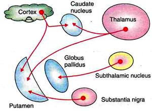
Fig.31. Afferent connections of corpus striatum.

Fig.32. Efferent and intrinsic connections of corpus striatum.
1. CONTROL OF VOLUNTARY MOTOR ACTIVITY
The movements during voluntary motor activity are initiated by cerebral cortex. However, these movements are controlled by basal ganglia, which are in close association with cerebral cortex. During lesions of basal ganglia, this controlling mechanism is lost and so movements become inaccurate and awkward. Basal ganglia control the motor activities because of the nervous (neuronal) circuits between basal ganglia and other parts of the brain involved in motor activity. The neuronal circuits arise from three areas of the cerebral cortex.
1. Premotor area,
2. Primary motor area and
3. Supplementary motor area
All these nerve fibers from cerebral cortex reach the caudate nucleus. From here, the fibers go to putamen. Some of fibers from cerebral cortex go directly to putamen also. Putamen sends fibers to globus pallidus. The fibers from here run towards the thalamus, subthalamic nucleus of Luys and substantia nigra. The subthalamic nucleus and substantia nigra are in turn, projected into thalamus. Now, the fibers from thalamus are projected back into the primary motor area and the other two motor areas, i.e. premotor area and supplementary motor area.
Fibers between cerebral cortex and caudate nucleus are concerned with regulation of conscious movements known as the cognitive control of activity. The cortical fibers reaching putamen are directly concerned with control of subconscious execution of some movements during performance of trained motor activities, i.e. skilled activities.
2. CONTROL OF MUSCLE TONE
Gamma motor neurons of spinal cord are responsible for maintaining the tone of muscles, which is important for posture. The tone of the muscle also depends on actions of muscle spindle. Gamma motor neurons, muscle spindle and muscle tone are all controlled by basal ganglia especially substantia nigra. In the lesion of basal ganglia, tone increases leading to rigidity.
3. CONTROL OF REFLEX MUSCULAR ACTIVITY
Some of the reflex muscular activities, particularly visual and labyrinthine reflexes are important in the maintenance of posture. Coordination and integration of impulses for these activities depend upon basal ganglia.
During lesion of basal ganglia, postural movements, especially visual and labyrinthine reflexes become abnormal. These abnormal movements are associated with rigidity. Rigidity is because of the loss of inhibitory influence from the cerebral cortex on spinal cord via basal gangliа
4. CONTROL OF AUTOMATIC ASSOCIATED MOVEMENTS
Automatic associated movements are the movements in body, which take place along with some motor activities. Examples are the swing of the arms while walking, appropriate facial expressions while talking or doing any work. Basal ganglia are responsible for these movements. The lesion in basal ganglia causes absence of these automatic associated movements, resulting in poverty of movements. Face without appropriate expressions while doing any work is called mask like face. Body without associated movements is called statue like body.
5. ROLE IN AROUSAL (EXCITIVE) MECHANISM
Globus pallidus and red nucleus are involved in arousal mechanism because of their connections with reticular formation. Extensive lesion in globus pallidus causes drowsiness, leading to sleep.
ROLE OF NEUROTRANSMITTERS IN THE FUNCTIONS OF BASAL GANGLIA
The functions of basal ganglia on motor activities are executed by some neurotransmitters released by nerve endings within basal ganglia. Following neurotransmitters are released in basal ganglia.
Dopamine: It is released by dopaminergic fibers from substantia nigra to corpus striatum (putamen and caudate nucleus - nigra strial fibers). The deficiency of dopamine leads to Parkinsonism.
Gamma aminobutyric acid (GABA): It is secreted by intrinsic fibers of corpus striatum and substantia nigra.
Acetylcholine: It is released by fibers from cerebral cortex to caudate nucleus and putamen.
Substance P and enkephalins: These are released by fibers from globus pallidus reaching substantia nigra.
Noradrenaline: This is secreted by the fibers between basal ganglia and reticular formation.
Among all these neurotransmitters, dopamine and GABA are inhibitory neurotransmitters. So, the dopaminergic fibers and the fibers releasing GABA are inhibitory fibers. All other transmitters possess excitatory function.
Дата добавления: 2018-02-15; просмотров: 3246; Мы поможем в написании вашей работы! |

Мы поможем в написании ваших работ!