Components and connections of functional divisions of cerebellum
| Division Components Afferent connections | Efferent connections | ||
| Vestibulo-cerebellum | Flocculonodular lobe (Nodulus and Flocculi) | Vestibulocerebellar tract | 1. Cerebello-vestibular tract 2. Fastigiobulbar tract |
| Spinocerebellum | Lingula, central lobe, culmen, lobulus simplex, declive, tuber, pyramid, uvula, paraflocculi and medial portions of cerebral hemispheres | 1. Dorsal spinocerebellar tract 2. Ventral spinocerebellar tract 3. Cuneocerebellar tract 4. Olivocerebellar tract 5. Pontocerebellar tract 6. Tectocerebellar tract 7. Trigeminocerebellar tract | 1. Fastigiobulbar tract 2. Cerebelloreticular tract 3. Cerebello-olivary tract |
| Corticocerebellum | Lateral portions of cerebral hemispheres | 1. Pontocerebellar tract 2. Olivocerebellar tract | 1. Dentatothalamic tract 2. Dentatorubral tract |
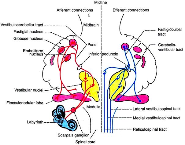
Fig. 26.
I. Cerebellovestibular Tract
Fibers of this tract arise from the flocculonodular lobe, pass through the inferior cerebellar peduncle of the same side and terminate on the vestibular nuclei in brainstem. The fibers from vestibular nuclei form medial and vestibulospinal tracts, which terminate on the medial group of alpha motor neuron in the spinal cord. This pathway forms the medial system of motor pathway (extrapyramidal system).
II. Dorsal Spinocerebellar Tract
This tract arises from Clarke's column of cells in the dorsal gray horn of spinal cord. It is uncrossed tract, and reaches the spinocerebellum through the inferior peduncle of same side. This tract conveys the proprioceptive information from the limbs of same side regarding the position and movements.
III. Ventral Spinocerebellar Tract
The fibers of this tract arise from the marginal cells in the dorsal gray horn of spinal cord. After taking the origin, the fibers cross the midline, ascend in the opposite side and reach the spinocerebellum through superior cerebellar peduncle. This tract conveys he information about the position and movements of opposite limbs to spinocerebellum.

Fig. 27. Connections of spinocerebellum.
IV. Olivocerebellar Tract
This tract is formed by the climbing fibers arising from the inferior olivary nucleus in medulla. After taking origin, these fibers cross the midline and reach the spinocerebellum through the inferior cerebellar peduncle of the opposite side. This tract also gives collaterals to the cerebellar nuclei particularly, the globosus nucleus and emboliform nucleus. The inferior olivary nucleus receives afferent fibers from three sources.
1. The brainstem nuclei of the same side
2. The spinal cord through spino-olivary tract of same side
3. Cerebral cortex of opposite side
The olivocerebellar tract conveys proprioceptive impulses from the body and output signals from cerebral cortex to spinocerebellum.
V. Pontocerebellar Tract
This tract arises from pontine nuclei, crosses the midline and reaches the spinocerebellum through the middle cerebellar peduncle of opposite side. The pontine nuclei receive afferents from cerebral cortex. The pontocerebellar tract conveys the information to spinocerebellum about the motor signals discharged from cerebral cortex.
VI. Tectocerebellar Tract
The tectocerebellar tract arises from the superior and inferior colliculi of tectum in midbrain. It reaches the spinocerebellum through the superior cerebellar peduncle of the same side. This tract carries visual impulses from superior colliculus and auditory impulses from inferior colliculus to spinocerebellum.
FUNCTIONS OF SPINOCEREBELLUM
Spinocerebellum regulates tone, posture and equilibrium by receiving impulses form proprioceptors in muscles, tendons and joints, tactile receptors, visual receptors and auditory receptors.
Spinocerebellum is the receiving area for the tactile, proprioceptive, auditory and visual impulses. It also receives the cortical impulses via pontine nuclei. The tactile and proprioceptive impulses are localized in the spinocerebellum. Localization of tactile and proprioceptive impulses in spinocerebellum is determined by stimulating the tactile receptors in skin and the proprioceptors and by recording the electrical responses in different parts of spinocerebellum. The different parts of the body are represented in the spinocerebellum in the following manner:
Lingula — Coccygeal region
Central lobe — Hindlimb
Culmen — Forelimb
Lobulus simplex — Face and head
Different parts of the body are represented in the cerebral cortex in an inverted manner. Whereas in the cerebellum the different parts are represented in upright manner.
Spinocerebellum regulates the postural reflexes by modifying muscle tone. It facilitates the gamma motor neurons in the spinal cord via cerebellovestibulospinal and cerebelloreticulospinal fibers. The gamma motor neurons reflexly modify the activity of alpha motor neurons and thus regulate the muscle tone. The lesion, destruction or abolishing the function of this part by cooling, causes stoppage of discharge from the gamma motor neurons resulting in hypotonia and disturbances in posture.
Spinocerebellum also receives impulses from optic and auditory pathway and helps in adjustment of posture and equilibrium in response to visual and auditory impulses.
Дата добавления: 2018-02-15; просмотров: 5457; Мы поможем в написании вашей работы! |

Мы поможем в написании ваших работ!