Mechanism of Saltatory Conduction
The myelin sheath is not permeable to ions. So, entry of sodium from extracellular fluid into nerve fiber occurs only in the node of Ranvier, where myelin sheath is absent. This causes depolarization in the node, and not in the internode. Thus, depolarization occurs at successive nodes. So, action potential jumps from one node to another. Hence, this is called saltatory conduction (saltare = jumping).
NERVOUS FIBERS CLASSIFICATION
1. Depending on structure:
a) Myelinated Nerve Fibers
Myelinated nerve fibers are covered by myelin sheath.
b) Non-myelinated Nerve Fibers
Nerve fibers of this type do not have myelin sheath.
2. Depending upon distribution:
а)Somatic Nerve Fibers - supply the skeletal muscles of the body.
b) Visceral or Autonomic Nerve Fibers - supply the various internal organs of the body.
3. Depending on source of origin:
a) Cranial Nerves - arising from the brain.
b) Spinal Nerves - arising from the spinal cord.
4. Depending on function:
а) Motor or Efferent Nerve Fibers - carry motor impulses from central nervous system to different parts of the body.
b) Sensory Nerve or Afferent Fibers - carry sensory impulses from different parts of the body to the central nervous system.
5. Depending on chemical neurotransmitter:
а) noradrenergic - secrete noradrenaline;
b) cholinergic - secrete acetylcholine.
6. Depending on diameter and conduction
| Fibers type | Fibers diameter (mcm) | Transduction velocity (m/sec) | Main function |
| Aα (Type I ) | 13-22 | 70-120 | skeletal muscles efferent fibers; receptors (muscular spindles) afferent fibers |
| Aβ (Type II ) | 8-13 | 40-70 | afferents from pressure and touching receptors |
| Aγ | 4-8 | 15-40 | receptors (muscular spindles) efferent fibers; part of afferents from pressure and touching receptors |
| Aδ (Type III) | 1-4 | 5-15 | afferents from skin temperature and pain receptors, partially pressure |
| B | 1-3 | 3-14 | autonomic nervous system preganglionar efferents |
| C (Type IV) | 0,5-1,5 | 0,5-2 | autonomic nervous system postganglionar efferents; pain and warmth skin receptors afferents |
Velocity of impulse through a nerve fiber is directly proportional to thickness of fibers. Except С type of fibers, all nerve fibers are myelinated, B type is partially myelinated.
NERVOUS FIBERS PROPERTIES
Excitability - nerve fibers have lower threshold for excitation than other cells. Resting membrane potential in nerve fiber is-70 mV. Firing level (critical depolarization level) is at-55 mV. Depolarization ends at +35 mV.
|
|
|
Conductivity - normally in body action potential is transmitted through nerve fiber in only one direction. However, in experimental conditions when the nerve is stimulated or damaged (tumor, anaesthesia, inflammation) action potential travels through the nerve fiber in both direction.
Refractory period
Summation. When one subliminal stimulus is applied, it does not produce any response in the nerve fiber. However, if two or more subliminal stimuli are applied within a short interval of about 0.5 m sec, the response is produced. This is because the subliminal stimuli are summed up together. This is known as summation.
Adaptation. While stimulating a nerve fiber continuously, excitability of nerve fiber is maximum at the beginning. Later - response decreases slowly and finally nerve fiber does not show any response at all. This phenomenon is known as adaptation or accommodation.
Infatigability. A nerve fiber cannot be fatigued, even if it is stimulated continuously for a long time. The reason for this is that nerve fiber can conduct only one action potential at a time. At that time, it is completely refractory and does not conduct another action potential.
Law “Everything or nothing”.
Excitement CONDUCTANCE through nervous fibers obeyes definite LAWS.
1) Physiological integrity law
tells that excitation conductance through nervous fibre is possible only in a case of its non-interrupted anatomical structure and physiological features.
2) Excitement conductance two-sided law
at irritation application on nervous fibre the excitement is diverged through it in both sides from irritation place (at tooth nerve irritation pain is stretched not only on local tissues but also irradiates in other body parts).
3) Excitement isolated conductance law
excitation through nervous fibers being in a composition of mixed nerves (for example, vagus) is diverged separately, i.e. it doesn’t transmit through one nervous fibre to another.
SYNAPSES PHYSIOLOGY
|
|
|
A synapse is a functional point of contact between 2 neurons, neuron and myocyte or neuron as well as neuron and secretory cell that transmits impulse from first to the second cell providing excitement (inhibiting) transmittance as well as nervous impulses transformation. The term “synapse” was introduced by English physiologist Charles Sherrington in 1897 who predisposed existence of special structural-functional formations providing contacts between neural cells.
TYPES:
1.According to communicational basis:
a) Central:
– Axo-somatic.
– Axo-dendritic.
– Axo-axonic.
– Dendro-somatic.
– Dendro-dendritic.
– Soma-somatic.
b) Peripheral:
– Neuro-muscular.
– Neuro-secretory.

Fig.12
2.According to nature:
– Electric synapses (ephapses).
– Chemical synapse.
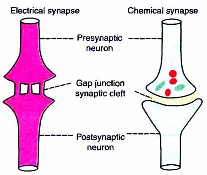
Fig.13
3. According to mediators – only chemical synapses.
4 main mediators groups:
a) acetylcholine;
a) catecholamines:
– dopamine;
– noradrenaline;
– adrenaline;
– serotonine (monoamine);
b) aminoacids:
– glycine;
– GABA (gama-amino-butiric acid);
– L-glutamate;
– cysteine and so on;
d) peptides.
4. According to ending effect:
– Stimulating – both electrical and chemical ones.
– Inhibiting- only chemical ones.
SYNAPSE STRUCTURE:
1.Presynaptic terminal or part – usually is presynaptic axon ending.
2.Synaptic cleft – is a space dividing membranes of contacting cells.
3.Post-synaptic part – is cell locus to which presynaptic ending comes.

Fig.14
Synaptic binding is interneuronal interaction main mechanism. It provides all major expressions of nervous system activity. It is one of the most essential structural-functional brain elements.
Neuron from which axon arises is called presynaptic neuron and neuron on which axon ends is called postsynaptic neuron. Axon of presynaptic neuron divides into many small branches before forming the synapse. These branches are known as presynaptic axon terminals. The slightly expanded presynaptic terminal has a definite intact membrane known as presynaptic membrane. Presynaptic terminal has two important structures:
|
|
|
1) mitochondria, which help in the synthesis of chemical neurotransmitter substances;
2) synaptic vesicles, which store neurotransmitter substances.
Membrane of postsynaptic neuron is called postsynaptic membrane. It contains some receptor proteins. Small space in between presynaptic membrane and postsynaptic membrane is called synaptic cleft. Basal lamina of this cleft contains cholinesterase, which destroys acetylcholine.
FUNCTIONS OF SYNAPSE
Main function of synapse is to transmit impulses, i.e. action potential from one neuron to another. However, some of synapses inhibit these impulses and so impulses are not transmitted to the postsynaptic neuron. Thus, synapses are of two types.
1. Excitatory synapses, which transmit the impulses-excitatory function and
2. Inhibitory synapses, which inhibit transmission of impulses—inhibitory function.
Дата добавления: 2018-02-15; просмотров: 1060; Мы поможем в написании вашей работы! |

Мы поможем в написании ваших работ!