Pre-auditory self-work materials. Basic knowledge, skills, experiences, necessary for study the topic: Subject To know To be able to Medical biophysics Muscular
Basic knowledge, skills, experiences, necessary for study the topic:
| Subject | To know | To be able to |
| Medical biophysics | Muscular contraction main and energetic parameters, difference between muscles stathic and dynamic work | Manage the methodics of force and work determining at muscular contraction as well as the one of useful action coefficient |
| Histology | General morphological-functional characteristics and histological structure of skeletal muscles | Manage the skills on skeletal muscles preparation identification |
| Biochemistry | Skeletal muscles biochemical structure and contraction fine biochemical mechanisms | |
| Sportive medicine | Representation about muscles force and work, their investigative methods (dynamometry, ergometry, ergography) | Assess and analize muscular force by dynamometry |
| Children diseases propedeutics | Skeletal muscles functioning peculiarities in a childhood | Investigate muscles tonus and force |
Topic content.
Muscles are classifiedby three different methods based on different factors.
1.Depending upon presence or absence of striations
2.Depending upon control and
3.Depending upon function
I. DEPENDING UPON STRIATIONS
Depending upon presence or absence of cross striations, the muscles are divided into two groups namely:
1. Striated muscle and
2. Non-striated muscle
Striated Muscle
Under light microscope, in each muscle cell, a large number of cross striations (transverse lines) are seen at regular interval. These muscles, having the cross striations are called striated muscles.
Skeletal muscle and .cardiac muscle belong to this category.
Non-striated Muscle
The muscles without cross striations are called non-striated muscles. These muscles are also known as plain muscles or smooth muscles.
II.DEPENDING UPON THE CONTROL
Depending upon control, the muscles are classified into two types namely:
1. Voluntary muscle and
2. Involuntary muscle
Voluntary Muscle
The activities of these muscles can be controlled voluntarily (at will). Skeletal muscles are the voluntary muscles. These muscles are innervated by somatic nerves.
Involuntary Muscle
The activities of these muscles cannot be controlled at will. Cardiac muscle and smooth muscle are involuntary muscles. These muscles are innervated by autonomic nerves.
III. DEPENDING UPON FUNCTION
The muscles are classified into three types depending upon function:
1. Skeletal muscle
2. Cardiac muscle and
3. Smooth muscle
Skeletal Muscle
Skeletal muscles are in association with bones forming the skeletal system. These muscles form from 40 to 50% of body mass. In human beings, about 600 muscles are identified. Skeletal muscles are voluntary and striated. These muscles are supplied by somatic nerves.
Striations, control and nerve supply of muscles
| Muscle | Striations | Control | Nerve supply |
| Skeletal muscle | Present | Voluntary | Somatic nerves |
| Cardiac muscle | Present | Involuntary | Autonomic nerves |
| Smooth muscle | Absent | Involuntary | Autonomic nerves |
Myofibrils or myofibrillae are fine parallel filaments present in sarcoplasm of the muscle cell. Myofibrils run through the entire length of the muscle fiber.
In cross section of a muscle fiber, the myofibrils are separated from one another by sarcoplasm. In some muscle fibers, some of the myofibrils are arranged in groups. These groups of myofibrils are called Cohnheim's areas or fields. The diameter of the myofibril is 0,2 to 2,0 microns. And, the length of a myofibril varies between 1,0 to 4,0 cm depending on length of the muscle fiber.
MICROSCOPIC STRUCTURE OF MYOFIBRIL
Light microscopic studies show that, each myofibril consists of a number of alternating light and dark bands. These bands are otherwise called the sections, segments or discs.
Dark band is called “A” band. “A” band is anisotropic. If polarized light is passed through the muscle fiber at this area, the light rays are refracted at different directions (An = not; iso = it; trops = turning). Light band is isotropic. Rays of polarized light, passed through the muscle fiber at this area, are refracted at the sail angle. So, this band is called “I” band.
The light band is otherwise called J band and the dark bands called Q disc (Querscheibe = cross disc).
In an intact muscle fiber, “I” band and “A” band of adjacent myofibrils are placed side by side. This gives the appearance of characteristic cross striations in muscular fiber.
A narrow lighter area called “H” zone (H = hell = light-in German, “H” = Henson - discoverer) is seen at the middle of “A” band. “I” band is divided into two by a narrow line called “Z” line (in German Zwischenscheibe = between discs). The portion of myofibril in between two “Z” lines is called sarcomere.

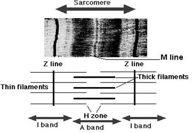
Fig.8. Structure of skeletal muscle.
SARCOMERE
Sarcomere is structural and functional unit of the skeletal muscle. Each sarcomere extends between two “Z” lines of myofibril. Thus, each myofibril contains many sarcomeres arranged in series throughout the length of myofibril.
Each myofibril consists of alternated dark “A” band and light “I” band (Fig. 8). In the middle of “A” band, there is a light area called “H” zone. In the middle of “H” zone middle part of myosin filament lies. It is called “M” line. Similarly, “'I” band is divided into two equal portions by means of a narrow line called “Z” line. Part of myofibril between two “Z” lines is sarcomere. When muscle is in relaxed state, the average length of each sarcomere is 2 to 3 microns.
COMPOSITION OF MUSCLE
Skeletal muscle is formed by 75% of water, 20% of proteins and 5% of organic substances other than proteins and some inorganic substances.
MUSCLE PROTEINS
Following are the proteins present in the muscle:
1.Myosin
2.Actin
3.Tropomyosin
4.Troponin
5.Actinin
6.Titin
7.Desmin
8.Myogen and
9.Myoglobulin.
Myogen is the protein present in sarcoplasm of the muscle cell. Myoglobulin is also present in sarcoplasm. This is also called myohemoglobin. Its function is similar to that of hemoglobin, that is to carry oxygen. This is a conjugated protein with molecular weight of 17,000.
Actinin attaches actin filament to “Z” line.
Titin is a large protein and it connects “M” line and “Z” line. Each titin molecule forms a scaffolding for sarcomere and provides elasticity to the muscle. When the muscle is stretched the titin unfolds itself. However, if stretching more it is offers resistance and protects sarcomere from overstretching.
Desmin binds “Z” line with sarcolemma.
SIMPLE MUSCLE CONTRACTION OR
TWITCH
The contractile property of muscle is studied by using the frog's gastrocnemius-sciatic preparation. This is also called muscle-nerve preparation.
When the stimulus with threshold strength is applied, the muscle contracts and then relaxes. These activities can be recorded graphically by using suitable instruments. The contraction is recorded as upward deflection from the base line. And relaxation is recorded as downward deflection back to the base line (Fig. 9).

Fig. 9. Isotonic simple muscle curve. PS = Point of stimulus. PC = Point of contraction. PMC = Point of maximum contraction. PMR = Point of maximum relaxation. LP = Latent period (0.01 sec). CP = Contraction period (0.04 sec). RP = Relaxation period (0.05 sec).
Simple contraction is called simple muscle twitch and graphical recording of this is called simple muscle curve. Four points are to be noted in this curve.
1. Point of stimulus (-PS): This denotes the time when the stimulus is applied.
2. Point of contraction (PC): This indicates the time when muscle begins to contract.
3. Point of maximum contraction (PMC): The muscle is contracting up to this point. This point also indicates the beginning of relaxation of the muscle.
4. Point of maximum relaxation (PMR): This point indicates complete relaxation of the muscle.
All these four points divide entire simple muscle curve into 3 periods.
1.Latent period (LP) is time interval between point of stimulus and point of contraction. There is no mechanical activity in the muscle during this period.
2.Contraction period (CP) is interval between point of contraction and point of maximum contraction. The muscle contracts during this period.
3.Relaxation period (RP) is the interval between point of maximum contraction and point of maximum relaxation. Relaxation of the muscle occurs during this period.
Duration of different periods in a typical simple muscle curve is as follows:
Latent period: 0,01 second
Contraction period: 0,04 second
Relaxation period: 0,05 second
Total twitch (contraction) period: 0,10 second
Contraction period is always shorter than relaxation period. This is because the contraction is active process and relaxation is passive process.
CONTRACTION TIME
Contraction time or total twich period in simple muscle varies from species to species. It is less in warm-blooded (homoiothermic) animals than in cold-blooded (poikilothermal) animals. In the same animal, it varies in different groups of muscles.
Based on contraction time, skeletal muscles are classified into two types, the red muscles and white muscles. Similarly, depending upon contraction time and myosin ATPase activity the muscle fibers are also divided into two types, type I and type II fibers. Type I fibers (slow fibers or slow twitch fibers) have small diameter. Type II fibers (fast fibers or fast twitch fibers) have large diameter. Most of skeletal muscles in human beings contain both types of fibers.
Red Muscles
Muscles containing large number of type I fibers are called red muscles, slow muscles or slow twitch muscles. These muscles have longer contraction time. Back muscles and gastrocnemius muscles are red muscles.
White Muscles
Muscles containing large number of type II fibers are called white muscles, pale muscles, fast muscles or fast twitch muscles. These muscles have shorter contraction time. Hand muscles and ocular muscles are white muscles.
The characteristic features of red and white muscles are given in table below.
FACTORS AFFECTING CONTRACTION FORCE
Skeletal muscle contraction force is affected by following factors:
A. Stimulus strength
B. Stimuli number
C. Temperature and
D. Load
A. Effect of Strength of Stimulus
If a series of electrical stimuli are applied by increasing the strength (voltage of current) each time, force of contraction is increased. Thus, the curves of different amplitude are obtained. The strength of stimuli is of five types.
1. Subminimal or subliminal stimulus: the muscle does not show any response.
2. Minimal stimulus: this is also called threshold or liminal stimulus - minimal contraction occurs.
3. Submaximal stimulus: muscle contraction force is increased.
4. Maximal stimulus: force of contraction reaches the maximum.
5. Supramaximal stimulus: beyond maximal strength, there is no further increase in contraction force.
Дата добавления: 2018-02-15; просмотров: 1914; Мы поможем в написании вашей работы! |

Мы поможем в написании ваших работ!