PERINATAL STATISTICS AND TERMINOLOGY
Table 1-5. Terminology for Perinatal Statistics

| Terminology | Definition |
| Gravidity | Total number of pregnancies irrespective of the pregnancy duration |
| Nulligravida | Woman who is not currently pregnant and has never been pregnant |
| Primigravida | Woman who is pregnant currently for the first time |
| Multigravida | Woman who is pregnant currently for more than the first time |
| Parity | Total number of pregnancies achieving ≥20 weeks’ gestation |
| Nullipara | Woman who has never carried a pregnancy achieving ≥20 weeks’ gestation |
| Primipara | Woman who has carried one pregnancy achieving ≥20 weeks’ gestation |
| Multipara | Woman who has carried more than one pregnancy to ≥20 weeks’ gestation |
| Parturient | Woman who is in labor |
| Puerpera | Woman who has just given birth |

12


| S2 OB-GYN.indb 12 | 7/8/13 6:35 PM | |||


 GI
GI

Chapter 1 l Reproductive Basics
Table 1-6. Terminology for Perinatal Losses

| Terminology | Definition |
| Abortion | Pregnancy loss prior to 20 menstrual weeks |
| Antepartum death | Fetal death between 20 menstrual weeks and onset of labor |
| Intrapartum death | Fetal death from onset of labor to birth |
| Fetal death | Fetal death between 20 menstrual weeks and birth |
| Perinatal death | Fetal/neonatal death from 20 menstrual weeks to 28 days after birth |
| Neonatal death | Newborn death between birth and the first 28 days of life |
| Infant death | Infant death between birth and first year of life |
| Maternal death | A woman who died during pregnancy or within 90 days of birth |
Table 1-7. Terminology for Mortality Rates

Terminology Definition
| Birth rate | Number of live births per 1,000 total population |
| Fertility rate | Number of live births per 1,000 women ages 15–45 years |
| Fetal mortality rate | Number of fetal deaths per 1,000 total births |
| Neonatal mortality rate | Number of neonatal deaths per 1,000 live births |
| Perinatal mortality rate | Number of fetal + neonatal deaths per 1,000 total births |
| Infant mortality rate | Number of infant deaths per 1,000 live births |
| Maternal mortality ratio | Number of maternal deaths per 100,000 live births |
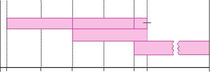


| Abortion | Fetal death | Neonatal death | ||||
| Perinatal death | ||||||
| Infant death | ||||||
| 0 2 | 10 | 20 | 30 | 40 | 1 month age | 1 year |
| Weeks of gestation/menstrual weeks | age | |||||
| Conception | Birth | |||||
| Figure I-1-4. Perinatal Mortality Terminology | ||||||


13
| S2 OB-GYN.indb 13 | 7/8/13 6:35 PM | |||

GI

USMLE Step 2 l Obstetrics
GENETIC DISORDERS

Human Genetics and Indications for Genetic Counseling
A 37-year-old G5 P0 Ab4 comes for prenatal care at 7 weeks’ gestation. She has experienced 4 previous spontaneous first-trimester abortions. She is concerned about the likelihood of her next pregnancy being successful.
• Advanced maternal age: women≥35 years of age at increased risk of fetal nondisjunc-tion trisomies (e.g., trisomies 21 and 18)
• Incidence of chromosomal abnormalities by maternal age:
| Age | Down Syndrome | Total Risk |
| 20 | 1 in 1,670 | 1:525 |
| 25 | 1 in 1,250 | 1:475 |
| 30 | 1 in 885 | 1:385 |
| 35 | 1 in 365 | 1:180 |
| 40 | 1 in 110 | 1:63 |
| 45 | 1 in 32 | 1:18 |
| 49 | 1 in 12 | 1:7 |

• Multiple fetal losses
• Previous child: neonatal death, mental retardation, aneuploidy, known genetic disorder
• Pregnancy or fetal losses: stillborn with birth defect, multiple pregnancy or fetal losses
• Family history: genetic diseases, birth defects, mental retardation
• Abnormal prenatal tests: triple marker screen, sonogram
• Parental aneuploidy
Chromosomal Aberration
Aneuploidy
This refers to numeric chromosome abnormalities in which cells do not contain 2 complete sets of 23 chromosomes. This usually occurs because of nondisjunction. The most common aneuploidy is trisomy, the presence of an extra chromosome. Most autosomal trisomies result in spontaneous abortions. The most common trisomy in first-trimester losses is trisomy 16. The most common trisomy at term is trisomy 21.
Polyploidy
This refers to numeric chromosome abnormalities in which cells contain complete sets of extra chromosomes. The most common polyploidy is triploidy with 69 chromosomes, followed by tetraploidy with 92 chromosomes. An example of triploidy is incomplete molar pregnancies,which occurs from fertilization of an egg by two sperm.
14

| S2 OB-GYN.indb 14 | 7/8/13 6:35 PM | |||
GI

Chapter 1 l Reproductive Basics
Structural alterations

This refers to conditions in which chromosomal material is deleted, gained, or rearranged. It can involve single or multiple chromosomes. An example of a chromosomal deletion is del (5p) or cri du chat syndrome, which is a deletion of the short arm of chromosome 5.
Mosaicism
This refers to the presence of >2 cytogenetically distinct cell lines in the same individual. Mosaicism can involve the placenta, the fetus, or both. Gonadal mosaicism can result in pre-mature ovarian failure and predispose the gonad to malignancy.
Common aneuploidies are as follows:

| Trisomy | Extra single | 47,XX+21 |
| Monosomy | Missing single | 45,X |
| Polyploidy | Extra set | 69,XXY |
Translocations
Reciprocal
This involves any two or more nonhomologous chromosomes, and occurs when there is a breakage and reunion of portions of the involved chromosomes to yield new products. Carriers of balanced reciprocal translocations have 46 chromosomes, with both derivative chromosomes present. The offspring may also have 46 chromosomes but have only one of the derivative chromosomes present.
Robertsonian
This always involves the acrocentric chromosomes, and is caused by centric fusion after loss of the satellite region of the short arms of the original acrosomic chromosome. The karyotype of a balanced Robertsonian translocation will appear to have only 45 chromosomes; however, the full complement of genetic material is present, and there are no clinical effects. The offspring may have 46 chromosomes but have double the genetic material of a particular chromosome.
Genetics of Pregnancy Loss
Miscarriage
At least 50% of first-trimester abortuses have abnormal chromosomes. The 2 most common aneuploidies in miscarriages are trisomy 16 and monosomy X. Fifty percent of these abnormali-ties are autosomal trisomies, with trisomy 16 being the most common.
15
| S2 OB-GYN.indb 15 | 7/8/13 6:35 PM | |||
GI
USMLE Step 2 l Obstetrics
OB Triad

Turner Syndrome
• Primary amenorrhea
• Web neck
• Streak gonads
OB Triad
Klinefelter Syndrome
• Testicular atrophy
• Gynecomastia
• Azoospermia
OB Triad
Down Syndrome
• Short stature
• Mental retardation
• Endocardial cushion cardiac defects
Turner Syndrome (45,X)

Also known as gonadal dysgenesis or monosomy X, Turner syndrome is seen in 1 in 2,000 births. In most cases it is the result of loss of the paternal X chromosome. Ninety-eight percent of these conceptions abort spontaneously. Obstetric ultrasound shows the characteristic nuchal skin-fold thickening and cystic hygroma. Those fetuses that survive to term have absence of secondary sexual development, short stature, streak gonads, primary amenorrhea, primary infertility, broad chest, and neck webbing. Urinary tract anomalies, bicuspid aortic valve, and aortic coarctation are commonly seen. Intelligence is usually normal. Mosaic patterns can occur with ovarian follicles present.
Дата добавления: 2018-11-24; просмотров: 253; Мы поможем в написании вашей работы! |

Мы поможем в написании ваших работ!